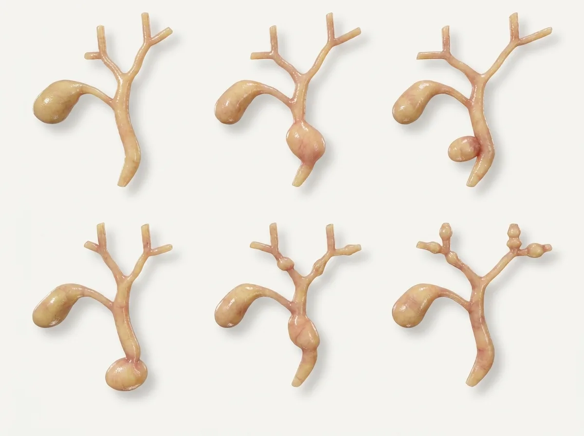
A choledochal cyst is an uncommon but important condition in which part of the bile duct becomes abnormally enlarged, forming a sac-like swelling. Although it is often thought of as a childhood disease, many patients in Bangladesh and across Asia are diagnosed only in adulthood, sometimes after years of unexplained abdominal pain, jaundice, or pancreatitis.
Choledochal cysts matter because, if left untreated, they can cause repeated infections, stones, pancreatitis, and, most importantly, a significantly increased risk of bile duct cancer. The good news is that complete surgical removal can prevent these problems. This article explains what choledochal cysts are, their types, symptoms, risks, and why complete excision by an experienced hepatobiliary surgeon is the recommended treatment.
Understanding the Bile Duct System
Bile is produced in the liver and travels through a network of tubes, the bile ducts, to reach the small intestine. Small ducts inside the liver join to form the right and left hepatic ducts, which unite to form the common hepatic duct. After the gallbladder's duct joins it, it becomes the common bile duct, which passes behind the duodenum and through the head of the pancreas. It meets the pancreatic duct and opens into the duodenum.
In a choledochal cyst, one or more parts of this duct system are abnormally widened (dilated), either as a round sac or a long, spindle-shaped swelling.
What Causes Choledochal Cysts?
The exact cause is not fully understood, but most cysts are believed to be congenital, meaning present from birth. The most widely accepted explanation is an abnormal junction between the bile duct and the pancreatic duct, called pancreaticobiliary maljunction. In this condition, the two ducts join outside the duodenal wall, forming an unusually long common channel. This allows pancreatic juice to flow back into the bile duct, where its powerful enzymes damage and weaken the duct wall over time, causing it to dilate.
Choledochal cysts are much more common in Asian populations than in Western countries and occur about three to four times more often in females than in males.
Types of Choledochal Cysts
Doctors classify choledochal cysts using the Todani classification, which helps decide the right operation:
Type I: the most common type, accounting for most cases. The common bile duct outside the liver is dilated, either as a round sac or a spindle shape.
Type II: a pouch (diverticulum) sticking out from the side of the bile duct. This type is rare.
Type III (choledochocele): a dilatation of the very end of the bile duct inside the wall of the duodenum.
Type IV-A: multiple cysts both inside and outside the liver. This is the second most common type.
Type IV-B: multiple cysts outside the liver only.
Type V (Caroli disease): multiple cysts of the bile ducts inside the liver only.
Symptoms in Children
In infants and children, choledochal cysts may cause:
Jaundice (yellowing of the eyes and skin), often coming and going
Pale, clay-coloured stools and dark urine
Abdominal pain
A lump or swelling in the upper right abdomen
Vomiting, fever, and poor feeding or poor weight gain
The combination of pain, jaundice, and an abdominal lump is called the "classic triad", though only a minority of patients have all three. Some cysts are now detected even before birth on routine pregnancy ultrasound.
Symptoms in Adults
In adults, symptoms are often vague, and the condition may be mistaken for gallstones or "gastric" problems. Common features include:
Recurrent pain in the upper abdomen
Episodes of jaundice
Cholangitis: fever with chills, jaundice, and pain caused by bile duct infection
Recurrent acute pancreatitis
Stones in the bile duct or inside the cyst
Sometimes, no symptoms at all, with the cyst found on a scan done for another reason
Complications If Left Untreated
Choledochal cysts tend to cause more problems over time. Possible complications include:
Recurrent cholangitis, which can become life-threatening
Stones forming inside the stagnant bile in the cyst
Recurrent pancreatitis
Secondary biliary cirrhosis: long-term liver damage from blocked bile flow
Cyst rupture, which is rare but serious
Bile duct cancer (cholangiocarcinoma) and gallbladder cancer: the most serious long-term risk
The Cancer Risk: Why Treatment Matters
People with choledochal cysts have a much higher risk of developing cancer of the bile duct and gallbladder than the general population. This risk increases with age: it is low in childhood but rises steadily in adulthood, particularly after the age of 30 to 40. The constant reflux of pancreatic juice and chronic inflammation are thought to drive cancer development in the cyst wall.
This is why the cyst should be completely removed, not just drained or bypassed. Older operations that simply connected the cyst to the intestine without removing it left the diseased lining in place, and many of these patients later developed cancer. Today, these "internal drainage" operations are no longer recommended.
How Are Choledochal Cysts Diagnosed?
Ultrasound: usually the first test, showing a dilated bile duct or cystic swelling
MRCP: the best non-invasive test. It clearly shows the type and extent of the cyst, the anatomy of the ducts, and the abnormal pancreaticobiliary junction.
CT scan: to assess complications and look for any signs of cancer
Blood tests: liver function, amylase and lipase, and tumour markers such as CA 19-9 and CEA in adults
ERCP: occasionally used for diagnosis or to treat infection or stones before surgery; also used for Type III cysts
Endoscopic ultrasound: in selected cases
Treatment: Complete Surgical Excision
The treatment depends on the type of cyst:
Type I and Type IV-B
The standard operation is complete excision of the cyst together with removal of the gallbladder, followed by reconstruction of bile flow using a Roux-en-Y hepaticojejunostomy. In this procedure, a loop of small intestine (jejunum) is brought up and joined directly to the healthy bile duct near the liver, so that bile drains freely into the intestine. The lower end of the cyst is removed down to where it meets the pancreatic duct, taking care not to injure the pancreatic duct.
Type II
The pouch is removed, and the bile duct is repaired. Sometimes a full excision with reconstruction is needed.
Type III
These are usually treated endoscopically by ERCP with a sphincterotomy (cutting the opening) or removal of the cyst through the endoscope. Surgery is rarely needed.
Type IV-A
The part of the cyst outside the liver is removed, with hepaticojejunostomy. If the cysts inside the liver are confined to one part of the liver, that part may also be removed (liver resection).
Type V (Caroli Disease)
If the disease is confined to one lobe of the liver, liver resection can be curative. When both lobes are affected and complications are severe, liver transplantation may be considered.
Open or Laparoscopic Surgery?
Choledochal cyst excision can be done by open surgery or, in experienced centres, by laparoscopic or robotic surgery. Minimally invasive surgery offers smaller scars and faster recovery, but it requires advanced skills because the hepaticojejunostomy is a delicate connection. The most important goals are complete removal of the cyst and a wide, tension-free connection that will drain well for life. The surgeon will choose the safest approach for each patient.
Recovery After Surgery
A hospital stay of about 5 to 10 days
A temporary abdominal drain in the first few days
Gradual return to oral feeding
Early walking to reduce the risk of chest infection and clots
Return to normal activities within 4 to 6 weeks
The removed cyst is examined in the laboratory to confirm there is no cancer
Possible Complications of Surgery
Bile leak from the new connection
Pancreatic leak, if the lower end of the cyst is close to the pancreas
Bleeding or infection
Narrowing (stricture) of the connection in the long term
Recurrent cholangitis or stone formation in a minority of patients
These risks are lowest when the operation is performed by a surgeon experienced in hepatobiliary reconstruction.
Long-Term Follow-Up
Even after successful surgery, a small risk of cancer in the remaining bile ducts persists, and the connection can narrow over many years. Long-term follow-up is therefore recommended:
Regular check-ups with liver function tests
Periodic ultrasound or MRCP
Prompt attention to any fever, jaundice, or abdominal pain
Choledochal Cysts in Pregnancy
Choledochal cysts are sometimes diagnosed for the first time during pregnancy, when the growing womb and hormonal changes can worsen symptoms. Management depends on the stage of pregnancy and severity of symptoms. In many cases, infections are controlled first, and definitive surgery is planned after delivery, with close cooperation between the surgeon and obstetrician.
Common Questions from Patients and Families
Is surgery needed even without symptoms?
Yes, usually. Even without symptoms, the cancer risk in the cyst wall increases with age, and surgery becomes more difficult once complications develop. Planned surgery after diagnosis is therefore the safer course.
Can surgery in a child be delayed?
For children, it is best not to delay long after diagnosis. Surgery at a younger age is associated with fewer complications and faster recovery.
Can I eat normally after surgery?
Yes. After recovery, most patients eat a completely normal diet and lead normal lives.
When to See a Specialist
A scan report mentioning a "dilated common bile duct" or "choledochal cyst"
Recurrent jaundice or cholangitis
Recurrent pancreatitis without an obvious cause
A child with jaundice, pale stools, or an abdominal lump
A history of a previous cyst drainage operation without removal
Conclusion
Choledochal cysts are uncommon, but they are not harmless. Without treatment, they can cause repeated infection, pancreatitis, and liver damage, and they carry a significant risk of bile duct cancer. Complete excision of the cyst with a well-performed Roux-en-Y hepaticojejunostomy is the standard, proven treatment and offers excellent long-term results.
If you or your child has been diagnosed with a choledochal cyst, or has an unexplained dilated bile duct, consult a hepatobiliary surgeon for a thorough evaluation and treatment plan.
This article is for general information and does not replace a personal medical consultation. Please discuss your individual treatment plan with your surgeon.