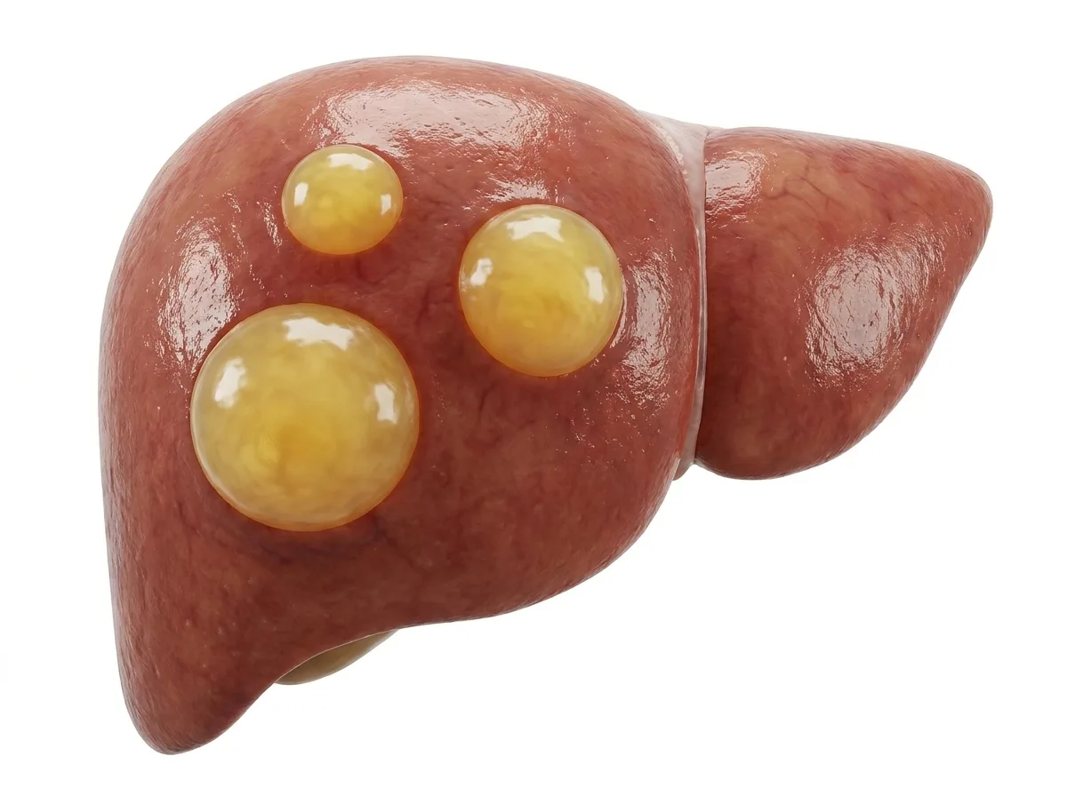
Few phrases cause as much anxiety as "a lesion was found in your liver." With the widespread use of ultrasound, CT, and MRI, liver cysts and masses are now discovered far more often, frequently by chance in people with no symptoms. The reassuring truth is that most liver lesions are benign (not cancer) and many need no treatment at all. However, some require careful monitoring, and a few need prompt surgery.
This article explains the common types of liver cysts and tumours, how doctors tell benign from malignant lesions, which tests are used, and when surgery or other treatment is recommended.
What Is a Liver Lesion?
"Lesion" is a general term for any abnormal area seen in the liver on a scan. It may be fluid-filled (a cyst) or solid (a mass or tumour). The word itself does not mean cancer. Identifying exactly what the lesion is, and whether it is harmful, is the job of the specialist.
Liver Cysts
Simple Liver Cysts
Simple cysts are thin-walled, fluid-filled sacs. They are very common, especially in women and older adults, and are almost always harmless. Most are small and cause no symptoms. They do not turn into cancer.
Treatment is needed only if a cyst becomes very large and causes pain, a feeling of fullness, or pressure on nearby organs. The preferred treatment is laparoscopic deroofing (fenestration), in which the outer wall of the cyst is removed so it cannot fill up again. Simply draining the fluid with a needle is not recommended on its own, because the cyst usually refills.
Hydatid Cysts
Hydatid disease is caused by the larval stage of a tapeworm, Echinococcus, which is carried by dogs. People become infected by swallowing eggs from contaminated food, water, or close contact with dogs. The cysts grow slowly over years and are often found in the liver.
Symptoms may include upper abdominal pain, a lump, or jaundice if the cyst ruptures into the bile ducts.
Rupture can cause severe allergic reactions or spread of the disease.
Treatment usually combines anti-parasitic medicine (albendazole) with surgery or, in selected cases, a needle-based procedure called PAIR (puncture, aspiration, injection, re-aspiration).
Surgery must be done carefully to avoid spilling the cyst contents.
Polycystic Liver Disease
An inherited condition in which many cysts develop throughout the liver, often along with kidney cysts. Most patients need no treatment, but a few with very large livers and severe symptoms may need surgery and, rarely, transplantation.
Mucinous Cystic Neoplasm of the Liver (Biliary Cystadenoma)
This is a rare cystic tumour, usually found in middle-aged women. It often has internal partitions (septations) or nodules on scans. Because it can turn cancerous (cystadenocarcinoma) and tends to recur if incompletely removed, complete surgical removal is recommended. Deroofing alone is not adequate.
Liver Abscess
Although not a tumour, a liver abscess is an important cause of a cystic lesion in Bangladesh. It is a collection of pus, caused by either bacteria (pyogenic abscess) or the parasite Entamoeba histolytica (amoebic abscess). Typical symptoms are fever, right upper abdominal pain, and weakness. Treatment involves antibiotics or anti-amoebic medicines, and often drainage by needle or tube under ultrasound guidance. Surgery is rarely required.
Benign Solid Liver Tumours
Haemangioma
The most common benign liver tumour. It is a cluster of blood vessels and is usually found by chance. Haemangiomas very rarely cause problems, do not turn into cancer, and almost never need treatment. Surgery is considered only for very large lesions causing symptoms.
Focal Nodular Hyperplasia (FNH)
The second most common benign tumour, most often seen in young women. FNH has a characteristic appearance on MRI, often with a central scar. It does not become cancerous and rarely bleeds, so it usually needs no treatment.
Hepatocellular Adenoma
A less common benign tumour, linked to long-term use of oral contraceptives, anabolic steroids, obesity, and some metabolic conditions. Unlike haemangioma and FNH, adenomas carry risks:
Bleeding or rupture, especially when larger than 5 cm
Transformation into cancer in a small proportion, particularly in men and certain subtypes
Management may include stopping hormonal medicines, weight loss, and close monitoring. Surgical removal is usually recommended for adenomas larger than 5 cm, for those that keep growing, for any adenoma in a man, and for certain high-risk subtypes.
Malignant Liver Tumours
Hepatocellular Carcinoma (HCC)
The most common primary liver cancer, usually developing in people with hepatitis B, hepatitis C, cirrhosis, or fatty liver disease. When detected early, it can be cured with surgical resection, ablation, or transplantation.
Intrahepatic Cholangiocarcinoma
A cancer arising from the bile ducts within the liver. It may cause vague pain, weight loss, or jaundice, but is often found late. Surgical resection with clear margins offers the best chance of cure, and chemotherapy is often added.
Liver Metastases (Secondary Liver Cancer)
Cancer that has spread to the liver from another organ is actually more common than primary liver cancer. The most common source is colorectal (bowel) cancer, followed by cancers of the pancreas, stomach, breast, lung, and neuroendocrine tumours.
Importantly, for patients with colorectal cancer that has spread only to the liver, liver resection combined with chemotherapy can offer long-term survival and even cure in a significant number of patients. Every such patient should be reviewed by a hepatobiliary surgeon.
Benign or Malignant? How Doctors Decide
Several clues help doctors decide whether a lesion is likely to be harmless or dangerous:
Patient background: hepatitis B or C, cirrhosis, or a history of cancer increase the concern.
Appearance on scans: many lesions have typical patterns on contrast CT or MRI that allow confident diagnosis without a biopsy.
Change over time: growth on repeat scans raises suspicion.
Tumour markers: AFP for HCC, and CEA and CA 19-9 for bile duct and metastatic cancers.
Symptoms such as weight loss, loss of appetite, or jaundice.
Diagnostic Tests
Ultrasound: the first test; it distinguishes cysts from solid lesions
Contrast-enhanced ultrasound: helpful in selected cases
Triple-phase CT scan: shows the blood-flow pattern of the lesion
MRI with liver-specific contrast: often the most accurate test for characterising lesions, especially FNH and adenoma
Blood tests: liver function, hepatitis markers, tumour markers, and hydatid serology if needed
Biopsy: used when the diagnosis remains uncertain after imaging. It is not routinely needed, and it is avoided in suspected hydatid cysts and in some potentially curable cancers.
PET-CT and colonoscopy: to look for a primary cancer when metastasis is suspected
Treatment Options
Observation
Many benign lesions, such as simple cysts, haemangiomas, and FNH, need no treatment. Sometimes a follow-up scan is recommended to confirm stability.
Liver Resection
Surgical removal of the part of the liver containing the lesion. It is used for malignant tumours, high-risk benign tumours such as large adenomas and mucinous cystic neoplasms, and symptomatic large benign lesions. Thanks to the liver's ability to regenerate, a large portion can be safely removed in a healthy liver. In selected patients, laparoscopic liver resection offers smaller scars and faster recovery.
Laparoscopic Deroofing
Used for large, symptomatic simple cysts.
Ablation
Heat-based destruction of small tumours using radiofrequency or microwave energy, especially for small HCCs and selected metastases.
Other Treatments
These include TACE, chemotherapy, targeted therapy, immunotherapy, and radiotherapy, depending on the tumour type and stage.
Recovery After Liver Surgery
After laparoscopic deroofing: usually home within 1 to 2 days
After liver resection: a hospital stay of about 5 to 10 days
Gradual return to normal activities over 4 to 8 weeks
Regular follow-up, with scans for malignant or high-risk lesions
Common Myths
"Every lump in the liver is cancer." Most liver lesions, especially in people without liver disease or cancer history, are benign.
"A cyst can be cured by draining it with a needle." Simple cysts usually refill after needle drainage alone; deroofing is the lasting treatment when treatment is needed.
"Haemangioma will turn into cancer." Haemangiomas do not become cancerous.
"If cancer has spread to the liver, nothing can be done." For colorectal and some other cancers, liver surgery combined with chemotherapy can offer long-term survival.
"Biopsy spreads cancer, so it should always be avoided." Biopsy is safe when indicated. Specialists choose when it is needed and when it can be avoided.
Accurate information helps patients avoid both unnecessary fear and dangerous delay.
Questions to Ask Your Doctor
What type of lesion do I have, and how sure are you of the diagnosis?
Is it benign or malignant, or is that still uncertain?
Does it need treatment, or can it be watched safely?
If watched, how often should I have scans?
If surgery is needed, can it be done laparoscopically?
Should I stop any medicines, such as oral contraceptives?
When to Seek Medical Attention
Any liver lesion found on a scan, especially if you have hepatitis B or C or cirrhosis
Sudden severe abdominal pain, which may suggest bleeding from a lesion
Fever with right upper abdominal pain
Jaundice, unexplained weight loss, or loss of appetite
A known lesion that is growing
Conclusion
Discovering a lesion in the liver can be frightening, but most liver cysts and tumours are benign. Many need no treatment, while others require monitoring or surgery. The key is an accurate diagnosis, usually achieved with high-quality imaging interpreted by an experienced specialist, so that harmless lesions are left alone and dangerous ones are treated promptly.
If a liver cyst or mass has been found on your scan, consult a hepatobiliary surgeon to understand what it is and whether any treatment is needed.
This article is for general information and does not replace a personal medical consultation. Please discuss your individual situation with your doctor.